Dracunculiasis
Overview
Dracunculiasis is infection with Dracunculus medinensis, a nematode worm (also known as roundworms) and mammalian parasite. It can also be referred to as Guinea Worm Disease (GWD). The female worms move through the subcutaneous tissue, causing intense pain, and eventually emerge through the skin, usually at the feet, producing oedema, a blister and eventually an ulcer, accompanied by fever, nausea, and vomiting. If they come into contact with water as they are emerging, the female worms discharge their larvae, setting in motion a new life cycle. GWD only occurs in the poorest 10% of world’s population who have no access to safe drinking water or health care. In 1991 there were 400,000 cases worldwide and in 2013 there were just 143, with hopes that it could soon become the first parasitic disease to be eradicated and the first disease to be eradicated without the use of vaccines or medical treatment.
Signs and symptoms
People with Guinea Worm Disease (GWD) are asymptomatic for 10-14 months while the worms breed and mature. Then, the person begins to feel ill as the female worm continues to grow and move down the muscle planes.
Initial signs and symptoms include:
-
Itchy rash
-
Slight fever
-
Nausea
-
Vomiting
-
Diarrhea
-
Dizziness
A blister then develops which can form anywhere on the skin. However,
the blister forms on the lower body parts in 80%–90% of cases. This
blister gets bigger over several days and causes a burning pain. When
the person puts this affected body part in cool water to ease the
symptoms, it is thought that the worm detects the temperature change and bursts from the blister to release hundreds of thousands of larvae into the water
In addition to the pain of the blister, removing the worm is also very painful. Furthermore, without proper care the wound often becomes infected by bacteria. These wound infections can then result in one or more of the following complications:
-
Redness and swelling of the skin (cellulitis)
-
Boils (abscesses)
-
Generalized infection (sepsis)
-
Joint infections (septic arthritis) that can cause the joints to lock and deform (contractures)
-
Lock jaw (tetanus)
If the worm breaks during removal it can cause intense inflammation as the remaining part of the dead worm starts to degrade inside the body. This causes more pain, swelling, and cellulitis.
While the death rate is low, disability is a common outcome of GWD. People have difficulty moving around because of pain and complications caused by secondary bacterial infections. The disability that occurs during worm removal and recovery prevents people from working in their fields, tending animals, going to school, and caring for their families. Disability lasts 8.5 weeks on average but sometimes can be permanent. When GWD was more common, the negative impacts on farming and livestock tending caused financial losses in the millions of dollars each year. In some villages where infection rates were high, more than 60% of children missed school. Some children were disabled by infection. Other children needed to work in place of disabled family members.
Causes
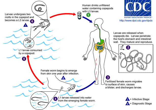
Dracunculiasis is caused by drinking water containing water fleas (Cyclops species) that have ingested Dracunculus larvae. Eggs/larvae require arthropods as intermediate hosts. Once ingested, copepods die and are digested to release the larvae. The larvae then penetrate the stomach or intestinal wall to enter the abdominal cavity or retroperitoneal space. Larvae mature into worms over 3 months, after which mating takes places and the male worm dies.
Approximately 1 year after mating, the fertilized females migrate through the subcutaneous tissues adjacent to long bones or joints towards the extremities. Once near the extremities they move towards the skin surface, resulting in blisters on the skin most commonly on the feet. The blister ruptures within 72 hours to expose one end of the emergent worm. The blister causes an extremely painful burning sensation as the worm emerges. The affected individual will often immerse the affected extremity in water to relieve the burning. When the blister is submerged in water, the adult female releases hundreds of thousands of guinea worm larvae and contaminates the water. The released larvae are eaten by copepods in the water and become infectious within 3 weeks to repeat the infectious cycle. Infected copepods can live in water for up to 4 months. Humans and dogs are the only known hosts to be infected.
Diagnosis / microbiology testing
Dracunculus medinensis is usually white with a spaghetti-like appearance. The female measures 1 metre in length and 1-2mm wide. The male worm is much smaller and rarely recovered from humans due to dying shortly after mating. GWD is usually diagnosed by visual examination when worm appears.
Microbiological tests are available to diagnose up to 6 months before emergence of worm but testing is expensive, complex and not well suited to the socioeconomically challenged areas in which Dracunculiasis still exists.
-
FAST-ELISA (Falcon Assay Screening Test-Enzyme-Linked Immunosorbent Assay)
-
EITB (Enzyme-linked Immunoelectrotransfer Blot)
Treatment
Management of GWD involves removing the whole worm and caring for the wound in general. There is no specific drug to treat or prevent GWD. There is also no vaccine to prevent GWD. The only way to avoid infection is to prevent exposure to the Guinea worm larvae in contaminated drinking water sources – boiling/chlorinating/sieving/education.
Optimal management of GWD involves the following steps:
-
First, each day the affected body part is immersed in a container of water to encourage more of the worm to come out. To prevent contamination, the infected person is not allowed to enter drinking water sources.
-
Next, the wound is cleaned.
-
Then, gentle traction is applied to the worm to slowly pull it out. Pulling stops when resistance is met to avoid breaking the worm. Because the worm can be as long as one meter in length, full extraction can take several days to weeks.
-
The worm is then wrapped around a rolled piece of gauze or a stick to maintain some tension on the worm and encourage more of the worm to emerge. This also prevents the worm from slipping back inside.
-
Afterwards, topical antibiotics are applied to the wound to prevent secondary bacterial infections.
-
The affected body part is then bandaged with fresh gauze to protect the site. Medicines, such as aspirin or ibuprofen, are given to help ease the pain of this process and reduce inflammation.
-
These steps are repeated every day until the whole worm is successfully pulled out
Tiabendazole and metronidazole have no effect on the worms but help reduce inflammation, enabling easier removal of worm.
Corticosteroid ointments shorten healing time and topical antibiotics may reduce risk of secondary bacterial infections.
Unerupted worms may be removed by minor surgery under local
anaesthesia.
Secondary bacterial infections should be treated with antibiotics.
Vaccines / preventative measures
There is currently no vaccine for GWD.
Preventative measures include protecting water sources and
filtering potentially contaminated water. The World Health
Organisation have provided endemic areas with drinking straws/
water filters to prevent ingestion of larvae.