Cysticercosis


Overview
Cysticercosis is a parasitic infection which affects tissues and skeletal muscles but can progress to the brain and central nervous system (known as neurocysticercosis). Less commonly, cysticercosis can localise in the skin, eyes and heart muscle. Serious disease is rare but can occur with neurocysticercosis or cardiac involvement. The causative organism is the larval cyst of the cestode, Taenia solium.
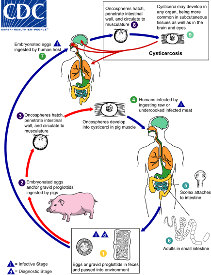
Cysticercosis is found worldwide, with the highest reported incidence of cases occurring in areas of Latin America, Asia and Africa where sanitation is poor. Cysticercosis is not to be confused with an intestinal tapeworm. Pork intestinal tapeworms are acquired when humans ingest larval cysts in raw or undercooked pork. Cysticercosis is acquired when humans ingest larval embryonated eggs, resulting in tissue infection with cysts.
Signs and symptoms
The time period between initial infection and the onset of symptoms can vary from several months to years. Infected individuals can remain asymptomatic even when harbouring multiple cysts throughout the body. Symptoms develop due to local inflammation at the site of infection and will depend upon the location, number, size and stage of the cyst. Cysts are also referred to as cysticerci and can be at a viable, degenerating or calcified stage. The intensity of the inflammatory response is a result of the degenerating stage.
Epilepsy is the most common manifestation and is present in 70-90% of symptomatic patients. Less frequent clinical manifestations include intracranial hypertension, hydrocephalus, chronic meningitis, and cranial nerve abnormalities. The most important clinical manifestations are caused by cysts in the central nervous system, known as neurocysticercosis. The resulting signs and symptoms depend on the number, location, size, and stage (viable, degenerating, or calcified) of the cysticerci and the intensity of the inflammatory response to any degenerating cysts.
Cysts in the brain or spinal cord cause the most serious form of the disease (neurocysticercosis), but can remain asymptomatic. Cysts are usually 5-20nm in diameter in brain parenchyma and up to 6cm in diameter in subarachnoid space. Cysts located within the brain ventricles can block the outflow of cerebrospinal fluid causing symptoms of intracranial pressure. Cysts involving the spinal cord cause symptoms of back pain and radiculopathy.
Neurocystericercosis signs and symptoms include:
-
Seizures
-
Headaches
-
Confusion
-
Difficulty with balance
-
Swelling of the brain
-
Excess fluid around the brain
-
Back pain
-
Radiculopathy
-
Stroke
-
Death
Cysts in the voluntary muscles are generally asymptomatic but can produce lumps under the skin which become tender.
Can result in the complications of:
-
myositis
-
fever
-
eosinophilia
-
muscular pseudohypertrophy
Cysts can also develop in the extraocular muscles and conjunctiva causing visual disturbances and difficulties, retinal oedema, haemorrhage and visual loss. Subcutaneous cysts present as firm, mobile nodules, most commonly occurring on the trunk and the extremities.
Causes
Infection is acquired by consumption of T.solium eggs
which have been excreted in the stool of another
individual with an adult tapeworm. This can occur when
an individual consumes food or drink contaminated with
eggs or puts contaminated fingers in their mouth.
An individual with an adult tapeworm is infected with taeniasis
and will shed T.solium eggs in their stools. Eggs released in the
stool are then consumed by a pig enabling them to develop into
larvae and form cysts (cysterici) in the pig’s muscles and
tissues. Infection with the cysts is defined as cysticercosis.
Humans who eat raw/undercooked infected pork will
consume the cysts. Larvae then emerge from the cysts in
the human gastrointestinal tract and develop into adult
tapeworms, completing the cycle.
Raw/undercooked pork that is not infected with T.solium
will not cause cysticercosis. Individuals with tapeworm
infections can infect themselves with T.solium eggs to then
develop cysticercosis via autoinfection transmission. These
individuals can also go on to infect others if they practise poor
hygiene and contaminate food/water that others will consume.
The number of cysticerci in the host can vary from 1 to >1,000. In
smaller numbers of cysticerci, the initial host tissue reaction is usually
minimal. As they develop, cysticercus affects surrounding tissue as a
slowly growing mass which can cause pressure atrophy. The majority of live
cysts do not cause an inflammatory reaction. An acute
inflammatory response occurs when cysts degenerate as
parasitic antigens are released. Degeneration of a cyst
can occur years after the initial infection.
At risk groups / risk factors
-
Individuals living within the same household as an infected person
-
Increasing age
-
Frequency of pork consumption
-
Poor household hygiene
Diagnosis / microbiology testing
-
Detection of calcified cysts on radiographs (asymptomatic patients)
-
CT or MRI scans show unilocular cysts (neurocysticercosis)
-
Positive ELISA indicates prior exposure to T.solium antigens
-
Immunoblotting techniques using purified glycoprotein from cyst-fluid may be more specific
-
Increased eosinophil number in CSF
-
Ophthalmic cysticercosis visualised through fundoscopy
Diagnosis often requires both imagining and serological testing due to:
-
Infection with very few cysticerci will show negative serum results
-
Cysterci may be in locations other than the brain so CNS imaging may be negative
-
Location and characteristics are best determined using MRI imaging
Treatment
Surgical removal is the treatment of choice for symptomatic cysts located outside of the central nervous system. Drug therapy for neurocysticercosis is controversial. Antihelminthic treatment causes the death of larvae which in turn triggers an inflammatory response that can exacerbate symptoms. Antihelminthic treatment should be avoided in cases where worms are dead (calcified cysts).
For complicated neurocysticercosis, treatment involves either
-
high-dose praziquantel 50mg/kg/day for 1-21 days OR
-
albendazole 10-15mg/kg/day for 7-30 days
Clinical evidence supports the use of albendazole over praziquantel and also suggests longer courses may be required for individuals with multiple cysts.
CNS inflammation can be managed with concurrent administration of dexamethasone but should be used in caution with praziquantel (reduced antihelminthic effect).
Seizures should be treated with anti-epileptic medication.
Continued treatment with anticonvulsants and other symptomatic medications may be required in cases where pathology is irreversible.
Vaccines / preventative measures
No human vaccine exists but studies have shown vaccinating pig intermediate hosts with T.solium antigens can cause disruption of the parasite life cycle.
Prevention is based on preventing the faecal-oral transmission of eggs from individuals infected with Taeniasis. Prevention methods include careful hand washing, avoiding placing hands in the mouth, avoiding eating from areas with poor hygiene practices and thoroughly cooking pork.

